KIT - Karlsruhe Institute of Technology
image/svg+xml
Institute for Applied Physics - Research Group Prof. Dr. Nienhaus
skip navigation
Home
Privacy Policy
Accessibility
KIT
de
Staff
Research
Start
Research
Research
Lebensprozesse in Zellen und Organismen
Protein-Nanopartikeln Wechselwirkung
Fluoreszierender Proteine
methods
Start
Research
methods
methods
FTIR spectroscopy
fluorescence fluctuation spectroscopy
single-molecule spectroscopy
high-resolution optical microscopy
atomic force microscopy
3D optical microscopy
Einzelmoleküluntersuchungen
Nanopartikelsynthese
Hämproteine
Lectures
Start
Lectures
Lectures
SS 2024
Vorlesungen/Seminaren-Inhalt
Start
Lectures
Vorlesungen/Seminaren-Inhalt
Vorlesungen/Seminaren-Inhalt
Biophysics
HS Nanoscopy
HS Conformational dynamics
WS 2023/24
SS 2023
WS 2022
SS 2022
WS 2021
SS 2021
WS 2021
SS 2020
WS 2020
SS 2019
WS 2018
SS 2018
WS 2017
SS 2017
WS 2016
SS 2016
WS 2015
SS 2015
WS 2014
SS 2014
WS 2013
SS 2013
SS 2012
Start
Lectures
SS 2012
SS 2012
Vorlesungsfolien
Skripte
Übungsblätte
WS 2011
Start
Lectures
WS 2011
WS 2011
Vorlesungsfolien
Skripte
Übungsblätte
SS 2011
Start
Lectures
SS 2011
SS 2011
Vorlesungsfolien
Skripte
Übungsblätte
WS 2010
Start
Lectures
WS 2010
WS 2010
Vorlesungsfolien
Skripte
Übungsblätte
SS 2010
Start
Lectures
SS 2010
SS 2010
Vorlesungsfolien
Skripte
Übungsblätte
WS 2009
Start
Lectures
WS 2009
WS 2009
Vorlesungsfolien
Skripte
Übungsblätte
Publications
Start
Publications
Publications
2024
2023
2022
2021
2020
2019
2018
2017
2016
2015
2014
2013
2012
2011
2010
2009
2008
2007
2006
2005
2004
2003
2002
2001
1997 - 2000
up to 1996
Seminars / Meetings
Start
Seminars / Meetings
Seminars / Meetings
Group seminar
Start
Seminars / Meetings
Group seminar
Group seminar
Past seminars
Meetings
Links
Job positions
How to find us
intern
Startpage
Home
Privacy Policy
Accessibility
Institute for Applied Physics - Research Group Prof. Dr. Nienhaus
Startpage
Research
Startseite
Staff
Research
Lectures
Publications
Seminars / Meetings
Links
Job positions
How to find us
intern
Research
Lebensprozesse in Zellen und Organismen
Protein-Nanopartikeln Wechselwirkung
Fluoreszierender Proteine
methods
Einzelmoleküluntersuchungen
Nanopartikelsynthese
Hämproteine
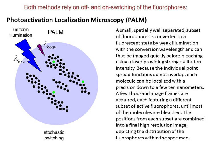
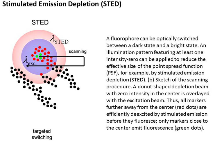
Super-resolution optical microscopy
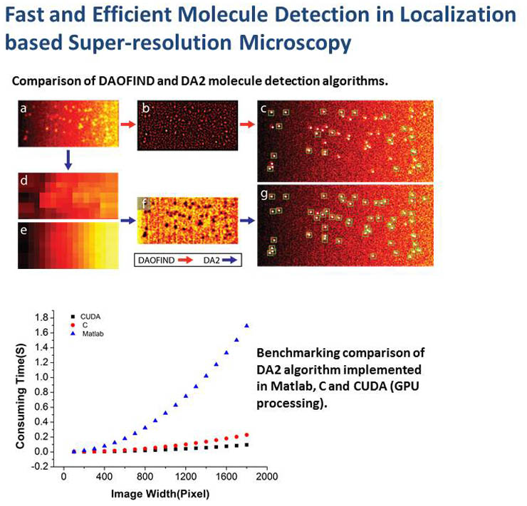
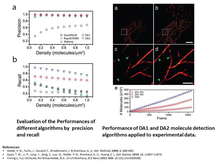
a-live PALM software
(Ref: Li et al, ACS Nano 2013): available upon request